martes, 27 de octubre de 2020


REMDESIVIR COMO TRATAMIENTO PARA COVID-19 - REPORTE FINAL

A pesar de que se han evaluado distintos agentes terapéuticos para el tratamiento de la enfermedad del coronavirus 2019 (Covid-19), no se ha comprobado la eficacia en agentes no antivirales.

¿Se podrá demostrar una eficacia significativa para combatir la enfermedad por coronavirus con remdesivir?

CONOCE EL ARTÍCULO DANDO CLICK AQUÍ

domingo, 2 de agosto de 2020


HIDROXICLOROQUINA CON O SIN AZITROMICINA EN COVID-19 LEVE A MODERADO

La hidroxicloroquina y la azitromicina han sido utilizadas para tratar pacientes con la enfermedad del coronavirus 2019 (Covid-19). Sin embargo, la evidencia en la seguridad y eficacia de éste tratamiento es limitado.
Se realizó un estudio multicéntrico, aleatorio, de ensayo abierto, 3 grupos controlados con pacientes hospitalizados en sospecha o confirmación de Covid-19. ¿Cuáles fueron las dosis usadas? ¿Qué efectos secundarios se presentaron? ¿El tratamiento combinado obtuvo mejor respuesta en los pacientes?

Da click AQUÍ para enterarte de ¡TODO!

sábado, 20 de febrero de 2016

Disculpas por el estancamiento del Blog

Buen día InterLector, comienzo esta nota pidiendo una GRAN disculpa al tener muy inactivo el sitio. La realidad es que entre el trabajo y los pacientes ha sido sumamente complicado mantenerme a la par de las publicaciones como previamente lo había estipulado, los tiempos son cortos y probablemente las fechas de publicación se cambien en un futuro.
Mi consciencia no me deja en paz y me siento a deuda con todos ustedes, los lectores que visitan asiduamente el lugar y aportan con ideas, comentarios y correcciones. Créanme que mantener un blog actualizado y al día es algo complicado y que involucra compromiso. Por esta razón he roto el hielo y he expuesto la razón de este estancamiento en el Blog del Internista, la buena noticia es que el "schedule" de las publicaciones cambiará y lo más probable es que sea los fines de semana.
Para no entrar en chismes y rumores, me despido de ustedes y les dejo las fechas probables de publicación a futuro. Los resúmenes se realizarán solamente 1 vez al mes, como es una recopilación de información de varios libros, bueno, me será imposible hacerlo 1 vez a la semana.


¡Pasen la voz InterLectores!

Rossyta Corb,
Admin del Blog del Intenista

Sterile Pyuria


Piuria estéril.

La piuria es definida como la presencia de 10 o más glóbulos blancos por milímetro cúbico en una muestra de orina, 3 o más glóbulos blancos por campo de alta potencia en orina centrifugada, un resultado positivo en la tinción de Gram en orina centrifugada o mediante una prueba de varilla graduada positiva para esterasa de leucocitos.  La piuria estéril es una condición prevalente y los estudios basados en la población general muestran que el 13.9% de las mujeres y 2.6% de los hombres son afectados.
Sin embargo, la pregunta clave es ¿Cuáles son las posibles causas? Y ¿Cómo se evalúa a un paciente con piuria estéril y cuáles son los posibles tratamientos?


Entérate en este artículo del NEJM, da click AQUÍ

viernes, 11 de diciembre de 2015

An Exophytic Mass on the Mandible of an Immunocompromised Man - ANSWER

Figure 1.

Figure 2.


Diagnosis: Coexistent Cryptococcus neoformans and Kaposi sarcoma in a patient with AIDS.


Histopathologic examination revealed multiple budding yeast forms surrounded by a clear halo (Figure 1A).Grocott-methaminesilver and mucicarmine (Figure 1B) stains highlighted these organisms and their capsules, respectively. A proliferation of atypical spindle cells arranged in fascicles, associated with slit-like vascular spaces and extravasated red blood cells, was also identified (Figure 1A). Immunohistochemistry for human herpesvirus 8 demonstrated positive nuclear staining within these spindled cells (Figure 2). These findings were diagnostic for cutaneous Cryptococcus infection in the context of Kaposi sarcoma (KS).
The patient was treated with amphotericin B and flucytosine followed by fluconazole for his cryptococcal infection; emtricitabine, tenofovir, and raltegravir were initiated as therapy for AIDS. Doxorubicin therapy was initiated for probable multifocal KS. The patient’s cutaneous lesions have improved on this regimen, with concomitant resolution of his lower extremity swelling and improvement in breath sounds. His human immunodeficiency virus (HIV) load is currently undetectable.
Cutaneous disorders are estimated to affect approximately 64% of patients with HIV, with an increasing prevalence at lower CD4 counts. These conditions include common infections and malignancies such as Staphylococcus aureus and squamous cell carcinoma, as well as a variety of inflammatory dermatoses that are often more severe than in immunocompetent patients. Of particular concern are those opportunistic infections and neoplasms that are classified as AIDS-defining illnesses, including cryptococcosis and KS. Often these conditions have a protean presentation and may simulate one another. Nevertheless, the coexistence of Cryptococcus and KS in a single clinical lesion is an uncommon occurrence. Colocalization of these infections may be the presenting sign of AIDS in patients with known HIV or those who had been previously undiagnosed; it has also been associated with paradoxical immune reconstitution inflammatory syndrome (IRIS) following initiation of highly active antiretroviral therapy (HAART)
The latter association is particularly noteworthy as significant morbidity and mortality are associated with KS-IRIS, particularly among patients with visceral KS.
KS-IRIS may be more difficult to treat than other forms of IRIS and has important prognostic implications. Cutaneous biopsy with meticulous histologic evaluation is therefore suggested in all HIV-infected patients with new or unusual skin lesions, even in the context of previously treated or active skin disorders. This practice guards against misdiagnosis of alternative or coincident disease. Multiorgan evaluation for cutaneous and visceral KS may be especially prudent in those patients who have also recently initiated HAART.
Rarely, KS can present with other viral, mycobacterial, or opportunistic fungal infections in the same lesion. In addition to cryptococcosis, other coincident infections include cytomegalovirus, molluscum, Candida albicans, Mycobacterium tuberculosis, Histoplasma capsulatum, and Mycobacterium aviumintracellulare. An instance of KS coexistent with both Cryptococcus and Mycobacterium avium-intracellulare has also been reported. The etiology of this phenomenon is unknown, and may represent a chance occurrence. It has alsobeen hypothesized that the vascular lesions of KS represent an ideal environment for the growth and protection of bloodborne opportunistic infections. 
Conversely, cryptococcal infection may induce a local inflammatory milieu that is hospitable to the development of KS, a concept known as inflammatory oncotaxis. These suppositions remain speculative, however, and the pathophysiologic mechanisms underpinning this unusual occurrence remain to be formally elucidated.


Clinical Infectious Diseases 2014;58(4):540
DOI: 10.1093/cid/cit711

A 59-Year-Old Woman With Chronic Skin Lesions of the Leg - ANSWER


Diagnosis: Acrodermatitis chronica atrophicans. 

The patient’s photograph of her skin rash taken 3 years ago, which clearly showed that she had erythema chronicum migrans (ECM) at that time, immediately prompted the diagnosis of Lyme disease in the form of acrodermatitis chronica atrophicans (ACA). The histology of the skin biopsy was compatible with this diagnosis, which was confirmed with serology and polymerase chain reaction (PCR). An enzyme-linked immunosorbent assay showed strongly elevated serum antibodies (immunoglobulin G [IgG]) to Borrelia burgdorferi (LiaisonDiaSorin; detection of immunoglobulin against B. burgdorferi, Borrelia afzelii, and Borrelia garinii; IgG >240 UA/mL). Western blotting (Biognost, Borrelia Euroline-WB) detected bands positive against VlsE, p83, p39, p30, and p21 antigens [1]. PCR on the skin biopsy sample (primer sets targeting 23S rDNA; TaqMan) was also positive. The patient was treated for 4 weeks with 100 mg of doxycycline twice a day. Six months later, she had no more lesions. Lyme borreliosis is caused by tick-transmitted spirochetes of the B. burgdorferi sensu lato complex. Although B. burgdorferi sensu stricto is the only species known to cause human disease in North America, at least 5 species can cause the disease in Europe: B. afzelii, B. garinii, B. burgdorferi sensu stricto, Borrelia spielmanii, and Borrelia bavariensis. The clinical symptoms vary widely and depend on the species; some have been described only in Europe [2]. 
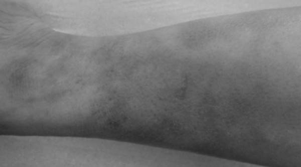
ACA appears to be due only to B. afzelii [3]. This dermatological entity is a rare tertiary manifestation of Lyme disease, manifesting as inflammatory and trophic lesions on acral skin. After an early inflammatory stage with bluish-red discoloration and doughy swelling of the skin, a late atrophic stage appears a few weeks or months later. The skin becomes thin, wrinkled, dry, and transparent because of the loss of epidermal and dermal structures. Vessels may be easily visible, and telangiectasias can be observed. The diagnosis is suggested by dermatologic lesions and a clinical history of tick bites or other well-defined manifestations of Lyme borreliosis, such as ECM, shown in our patient’s picture. Confirmation of the diagnosis is obtained by serological testing (enzyme immunoassay and Western blotting). These methods might increase diagnostic accuracy over that of PCR, which has a sensitivity of about 50%, depending on primer set [4]. Treatment of ACA is usually based on a course of antibiotic treatment with ceftriaxone [5] or doxycycline [6] for 21–28 days. Complete disappearance of lesions is normally described [7, 8]. The absence of treatment can lead to fibrotic nodules and/or patchy or bandlike indurations that may limit joint movement without treatment.


Clinical Infectious Diseases 2013;57(12):1782
DOI: 10.1093/cid/cit667


sábado, 19 de septiembre de 2015

A 59-Year-Old Woman With Chronic Skin Lesions of the Leg - QUIZ

Figure 1. Progressive atrophic and erythematous skin lesions on the left leg.

Figure 3. Asymptomatic skin rash on the back of the patient’s left ankle in July 2008.

Figure 2. Erythema with mild atrophy of the left foot.


A 59-year-old woman, a retired school director with no particular medical history, sought consultation because of chronic skin lesions on her left leg (Figures 1 and 2), which had been present for several months. Erythematous and minor atrophic lesions of the skin were seen. No infiltration was noted, and no other local symptom mentioned. The patient did describe pain in several joints. The physical examination found no other abnormalities. The patient also mentioned a history of a skin rash 3 years earlier, on the left ankle, which had appeared after a walk in a forest in southern France. A photograph she had taken then showed a large erythematous ring, which subsequently spread to the entire leg (Figure 3). No macular lesion was seen, and no pain at the time was reported. Laboratory analysis revealed a white blood cell count of 4420 cells/µL. Her C-reactive protein level was 3 mg/L (reference, <5 mg/L), and her fibrinogen level was 3 g/L (reference range, 2–4 g/L). Her liver enzyme and total complement activity levels were normal. The test for rheumatoid factor test was negative, but that for antinuclear antibodies was positive (1:200), with no specificity. The skin biopsy of a nonatrophic area showed cutaneous lymphoplasmacytic infiltration of interstitial tissue. 

What is your diagnosis?


Clinical Infectious Diseases 2013;57(12):1751
DOI: 10.1093/cid/cit661