


.png "Google-Translate-Spanish to English")


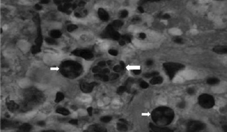 |
| Figure 1. Skin biopsy showing several endospores acattered throughout the dermis (small arrows) with a tightly packed, morula-like sporangium (large arrow, hematoxylin-eosin stain, magnification x 400). |
Diagnosis: Protothecosis.
Skin biopsy revealed several endospores (small arrows) scattered throughout the dermis with a tightly packed, morula-like sporangium (Figure 1). The fungal culture of the tissue confirmed the diagnosis of protothecosis. The patient was treated successfully with parenteral amphoteracin B followed by oral itraconazole.
Prototheca is a type of green alga found worldwide in sewage, fresh water, trees, and soil. Infection is usually caused by Prototheca wickerhamii. Less commonly, infection occurs with Prototheca zopfii. Infection is quite rare despite frequent exposure. Patients are typically immunocompromised with a
history of traumatic inoculation. The initial cutaneous lesion is typically a single localized eczematous nodule or plaque. An initial presentation with large extensive plaques or ulcers on bilateral forearms can be misleading for clinicians. Systemic dissemination may rarely occur. Given that the infection is quite rare, there is currently no evidence-based protocol for the management of protothecosis. However, there are case reports in the literature indicating that the use of intravenous amphotericin B is effective in disseminated disease.
Clinical Infectious Diseases 2013;56(2):307
0 comentarios:
Dí lo que piensas...